The body’s command center, the central nervous system (CNS) coordinates and controls several bodily activities and processes. The central nervous system comprises the brain and spinal cord functioning collaboratively to analyze information, initiate actions and regulate internal balance (Homeostasis). It is essential for our perception and interaction with the environment as well as for controlling movement, processing emotions and maintaining bodily functions in a regulated manner.
The brain, a complex and highly specialized organ is the command center of the CNS. One interesting fact about the brain is its remarkable energy consumption. Although the brain accounts for a mere 2% of our overall body weight, it utilizes approximately 20% of the body’s total energy resources. To function at its best, the brain needs regular doses of oxygen and nutrition.

Neuroplasticity, also referred to as the brain’s plasticity, is another fascinating characteristic of the brain. It is the term used to describe the brain’s capacity to undergo changes and adapt as a result of experiences, learning and external influences throughout an individual’s lifetime. It allows new connections to form between neurons, rewire neural pathways and recover from brain injuries. The spinal cord on the other hand serves as the conduit between body and the brain.
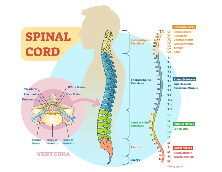
Did you know that the spinal cord has millions of nerve fibers and measures approximately 18 inches long? Remarkably, it can transmit signals to the brain at a remarkable speed of up to 268 miles per hour! It is protected by the vertebral column and spans from the base of the brain to the lower back. The spinal cord is also essential for controlling autonomic processes like digestion, blood pressure control and heart rhythm.
The brain and spinal cord work together as part of the central nervous system, synergistically ensuring the body’s optimal functioning. They are protected by layers of membranes called meninges and encased within the skull and vertebral column. A central nervous system disorder or injury can have a substantial impact on one’s general health and well-being.
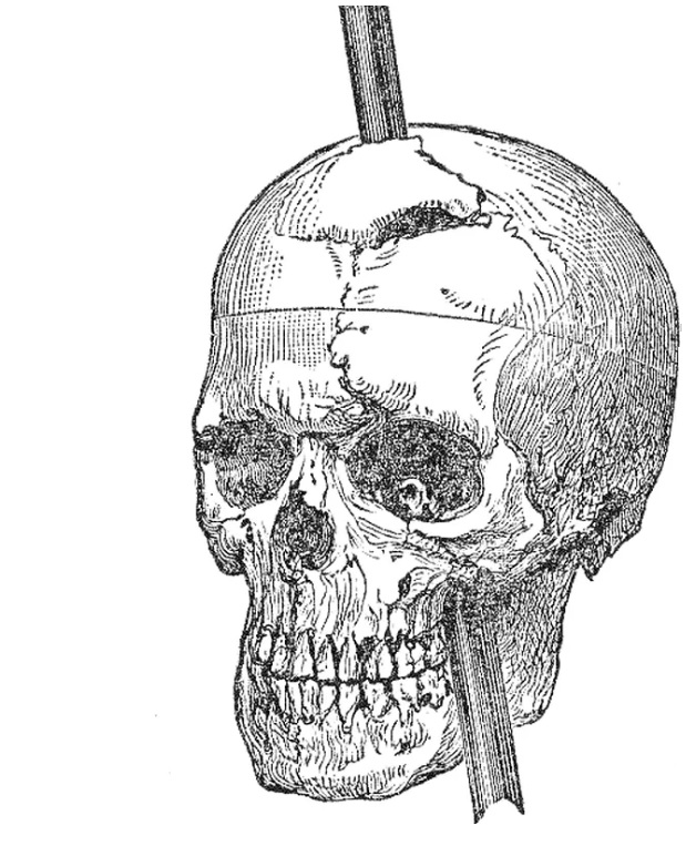
A Curious Case of Phineas Gage – A Traumatic Brain Injury Survivor
In the world of neuroscience, Phineus Gage is a well-known instance that demonstrates the extraordinary relationship between the brain and behavior. In 1848, Gage experienced a severe traumatic brain injury when an iron rod accidentally penetrated his skull damaging parts of his frontal lobe. What makes Gage’s case so intriguing is the significant change in his personality and behavior following the injury. Before the accident, he was described as responsible, hardworking and wee-mannered. His friends and family did however detect a significant change in his character after the accident. He became impulsive, irritable and exhibited socially inappropriate behavior.
This case shed light on the role of the frontal lobe in regulating personality and executive functions. Decision-making, impulse control and emotional regulation are all regulated by the frontal lobe. Gage’s injury provided evidence that damage to this area can have profound effects on one’s behavior and social functioning. This is why individuals with front-temporal dementia often experience initial symptoms related to changes in their personalities.

Exploring the Marvels of the Human Brain
An adult human brain typically weighs 3 pounds (1.4 kg). The brain which is relatively small in size but contains about 86 billion neurons, is a hub of activity. The transmission of electrical signals and the processing of information are made possible by the complicated network that connects these neurons.
Our capacity to adapt and learn in addition to our intelligence and consciousness are all functions of the brain. The brain is thought to be capable of storing up to 2.5 petabytes of data or which is approximately 3 million hours of television.
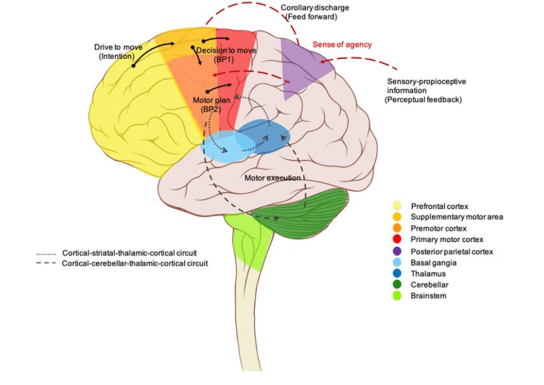
One of the key functions of the brain is cognition which allows us to perceive and understand the world around us, process information and engage in complex mental tasks. Different regions of the brain such as the amygdala and prefrontal cortex help recognize and respond to various emotional stimuli influencing our mood, behavior and social interactions. The motor cortex and other motor-related areas in the brain coordinate muscle movements allowing us to walk, talk and perform skilled tasks. The brain communicates with the spinal cord and peripheral nerves to control our body’s muscles.
Additionally, the brain plays a crucial role in overseeing fundamental bodily functions and upholding homeostasis. For instance, the hypothalamus is crucial for controlling hormone production, body temperature, hunger, thirst and sleep-wake cycles. It acts as a central command center receiving and sending signals to various parts of the body to maintain balance and ensure optimal functioning.
Additionally, the brain is involved in sensory perception allowing us to see, hear, taste, smell and touch. Different regions of the brain such as the visual cortex, auditory cortex and somatosensory cortex process and interpret sensory information enabling us to perceive our environment and interact with it. With billions of neurons that communicate by chemical and electrical signals, the brain is an extremely interconnected organ. It is organized into several areas and structures each with unique functionalities. These include the cerebral cortex, cerebellum, brainstem and various subcortical structures.
Statistics demonstrate that the human brain is prone to numerous disorders and diseases underscoring its susceptibility. Numerous neurological ailments including Parkinson’s disease, Alzheimer’s disease, stroke and epilepsy afflict millions of people worldwide according to the World Health Organization. To advance research and develop effective treatments for various illnesses, it is essential to comprehend the complexity of the human brain.
The Crucial Functions of the Spinal Cord
Nerve fibers are sensory neurons carrying impulses from various body parts to the spinal cord, where they are transmitted to the brain for interpretation and processing. This allows us to perceive and interpret sensations such as touch, temperature, pain and proprioception (awareness of body position and movement).
Apart from transmitting sensory information, the spinal cord has a critical function in controlling motor activities. Motor neurons within the spinal cord receive commands from the brain and transmit them to the muscles enabling voluntary movements. These actions can range from the simple to the complex such as walking or playing an instrument.
Another important function of the spinal cord is reflex action. Reflex is automatic, involuntary responses to specific stimuli that help protect the body from harm or maintain balance. When a sensory signal triggers a reflex such as touching a hot surface, the spinal cord initiates an immediate motor response without the involvement of the brain. This quick and automatic reaction helps prevent injury and allows for swift responses.

The spinal cord also plays a role in autonomic processes which are crucial for controlling unconscious body functions like respiration, blood pressure, digestion and heart rate. Nerve fibers in the spinal cord control the autonomic nervous system, which maintains homeostasis and ensures the proper functioning of internal organs.
Statistics show that spinal cord injuries affect millions of people worldwide. Around 17,810 new spinal cord injuries are reported annually in the United States alone according to the National Spinal cord Injury Statistical Center. These injuries can have a profound impact on individuals causing paralysis and loss of function.
To diagnose and treat spinal cord illnesses and injuries, it is essential to have a thorough understanding of the spinal cords functioning. Current research into spinal cord injuries aims to develop new therapies and rehabilitation techniques that will improve the quality of life for people with spinal cord injuries.
Development of The Central Nervous System
The development of the central nervous system (CNS) is a complex and intricate process that begins early in embryonic development and continues throughout life.
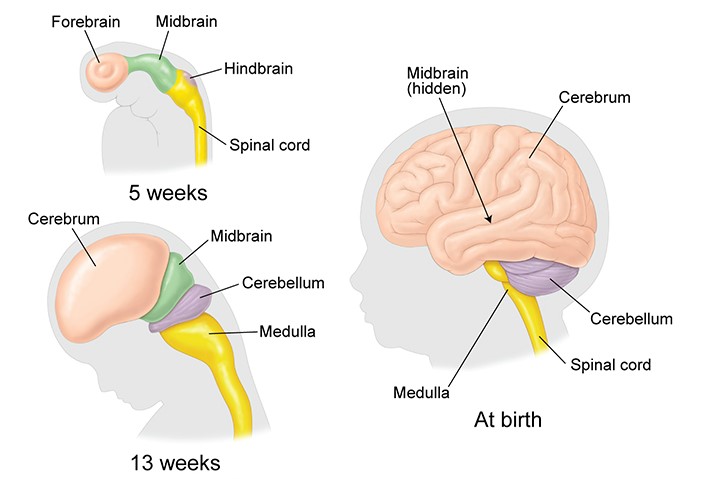
The beginning of the central nervous system (CNS) development, which eventually results in the development of the brain and spinal cord, is marked by the formation of the neural tube. The neural tube develops from a flat sheet of cells that folds and closes to form a hollow tube. This process known as neurulation occurs during the early stages of embryonic development.
As the neural tube develops, it undergoes further specialization. Neural progenitor cells divide within the neural tube to produce a variety of neurons and glial cells necessary for the healthy operation of the nervous system. Neurons carry out the transmission of electrical signals, whereas glial cells provide support and insulation to the neurons.
As the central nervous system (CNS) develops, various portions of the brain and spinal cord emerge, each with a distinct structure and function.
The forebrain, midbrain and hindbrain are some of these areas and they eventually give rise to specialized structures including the cerebral cortex, thalamus, hypothalamus, cerebellum and brainstem.

The CNS is affected by a variety of genetic and environmental variables during development. The wiring and function of the developing CNS can be shaped by environmental factors like nutrition, exposure to toxins and sensory experiences while genetic factors set the basic framework for CNS development.
The CNS continues to develop after birth and into early childhood as connections between neurons are improved and reinforced through a process known as synaptic pruning. The neuronal pathways can be adjusted throughout this procedure which also improves cognitive and motor skills. Our brain and spinal cord’s structural underpinnings are shaped by the extraordinary process of the central nervous system’s (CNS) development.
Some Fascinating Statistics about the Incredible Journey of CNS
Development
- Early formation: Did you know that three weeks after conception, the CNS starts to develop? The neural tube begins to take shape throughout the early stages and eventually develops into the brain and spinal cord.
- Rapid growth: During embryonic development, the CNS experiences rapid growth. In a short span of one month, the neural tube undergoes expansion and differentiation giving rise to distinct regions such as the forebrain, midbrain ad hindbrain.
- Neuronal connections: Neurons begin to link to one another as the CNS grows, creating complex networks that enable communication throughout the body.
- Synapse Formation: Synapses, the connections between neurons, are crucial for the CNS’s ability to convey information. Unbelievably, it is thought that a single neuron can link with thousands of other neurons forming a large and complex brain network.
- Neuroplasticity: During development, the CNS is extraordinarily elastic, enabling adaptability and rewiring in response to experiences and outside stimuli. During Childhood, when the brain experiences enormous changes and creates neuronal connections based on learning and development, this flexibility is most noticeable.
- Disorders and challenges: Unfortunately, the development of the CNS can be affected by various factors leading to developmental disorders and challenges. By statistics, 15 % of children worldwide suffer due to a neurodevelopmental difficulty such as attention-deficit/hyperactivity disorder (ADHD) or autism spectrum disorder.
- Lifelong Learning: Our central nervous system (CNS) develops during our lives as we continue to learn and acquire new knowledge. Neurogenesis and neuroplasticity are the terms used to describe this constant process of neuronal growth and adaptation which demonstrates the brain’s extraordinary capacity for development.
Relationship between the Central Nervous System and the Endocrine System
Even though they serve different purposes, they work together to maintain overall homeostasis and control a variety of body activities.
The endocrine system is made up of hormone-producing glands that control biological activity whereas the central nervous system (CNS) promotes neuronal connection and signal transmission by releasing neurotransmitters. These hormones function as chemical messengers that reach their intended cells and organs through the bloodstream. They control functions like reproduction, growth, metabolism and stress response.
To keep the body in balance and coordinate its different functions, the central nervous system’s (CNS) and endocrine system’s connection is crucial. This connection depends on the hypothalamus, a region of the brain. The pituitary gland, which releases hormones, is frequently referred to as the “master gland” due to its regulatory function over other endocrine glands, acting as a bridge between the CNS and the endocrine system.
Central Nervous System and its Interaction with the Immune System
The central nervous system (CNS) and the immune system are two interconnected systems in the human body that work closely together to maintain overall health and well-being. Let’s explore some fascinating statistics that highlight the intricate relationship between the CNS and the immune system.
- Communication Pathways: A network of cells, chemicals and molecular signals connects the CNS and immune system. The human brain is thought to have approximately 100 billion neurons and these send and receive signals that assist in controlling immunological reactions.
- Neuroinflammation: Immune responses may be significantly impacted by inflammatory events in the CNS. Numerous neurological conditions are influenced by neuroinflammation or inflammation of the brain and spinal cord. Data shows that neuroinflammatory illnesses like multiple sclerosis have an estimated incidence of about 2.5 million cases worldwide and pose a major negative influence on a large number of people.
- Neuroimmune Disorders: The interaction between the CNS and the immune system can be disrupted in certain disorders. Autoimmune conditions like Guillian-Barre syndrome and autoimmune encephalitis involve immune responses targeting components of the nervous system. A large number of people are impacted by these conditions and Guillian-Barre syndrome alone is estimated to affect about 1-2 people per 100,000 people annually.
- Stress and Immunity: Through the release of stress hormones, the CNS has the power to affect immunological responses. Chronic stress has been related to lowered immune function, increased susceptibility to infections and other immunological-related diseases. An estimated 20% of people worldwide are thought to suffer from stress-related ailments.
- Neuroimmune Interactions in Disease: The close interaction between the CNS and the immune system is evident in various neurological disorders. Immune reactions and inflammation in the brain are factors in neurogenerative illnesses including Alzheimer’s and Parkinson’s. These diseases have a significant impact on the quality of life with Alzheimer’s disease alone affecting more than 50 million individuals worldwide.
Understanding the CNS’s Control Over Voluntary Muscle Movements
Did you know that our bodies are composed of different types of muscle fibers? Fast-twitch fibers are responsible for explosive movements, like sprinting, while slow-twitch fibers are involved in endurance activities, such as long-distance running. The CNS coordinates the activation and recruitment of these fibers based on the specific demands of the movement. Also, ever wondered how athletes can execute complex movements effortlessly? Muscle memory, controlled by the CNS, plays a crucial role. With practice, the CNS establishes neural patterns that streamline movements, reducing the need for conscious effort and enhancing performance. Let’s delve into the fascinating mechanisms of how the CNS regulates and coordinates these movements.
- Motor Cortex: The frontal lobe of the brain’s primary cortex is in charge of organizing and carrying out voluntary muscular actions.
- Pyramidal System: The pyramidal system, consisting of the corticospinal and corticobulbar tracts, is a major pathway through which motor commands from the motor cortex reach the muscles. These tracts transmit signals from the motor cortex to the spinal cord (corticospinal) and brainstem (corticobulbar), where they synapse with lower motor neurons that directly control muscle contractions.
- Basal ganglia: The basal ganglia, a collection of structures deep within the brain are involved in motor control and play a role in refining and fine-tuning voluntary movements.
- Cerebellum: The cerebellum can regulate and modify motions to ensure correctness, precision and smoothness because it receives input from sensory systems, motor parts of the brain and feedback from muscles.
- Feedback Mechanisms: Sensory receptors in muscles, tendons and joints provide information about the positron, tension and movement of the body parts. This feedback helps the CNS continuously modify muscle contractions to maintain balance, posture and control.
- Motor Learning: With practice, the CNS establishes new neural connections, optimizing the efficiency and accuracy of movements. This process allows us to learn and perform complex tasks such as playing musical instruments or sports.

Understanding the Structure and Function of Neurons
- Structure: Each neuron consists of three main parts; the cell body, dendrites and axon. The cell body contains the nucleus and other vital components necessary for the neuron’s survival and functioning. Dendrites are branching extensions that receive signals from other neurons and transmit them to the cell body. The axon is a long, slender projection that carries signals away from the cell body and transmits them to other neurons or target cells.
- Function: Action potentials or electrical signals are vital for moving throughout the nervous system and are transmitted by neurons. These signals allow for rapid communication and coordination of various bodily functions. Through its dendrites, a neuron receives signals from other neurons, integrates the data and then produces an electrical impulse that passes down its axon. At the end of the axon, specialized structures called synapses allow the neuron to pass the electrical signal to the next neuron or target cell, such as a muscle or gland.


